References

Agustí A, Celli BR, Criner GJ Global initiative for chronic obstructive lung disease 2023 report: GOLD executive summary. Am J Respir Crit Care Med. 2023; 207:(7)819-837 https://doi.org/10.1164/rccm.202301-0106PP

Celli BR, Cote CG, Marin JM The Body-Mass Index, Airflow Obstruction, Dyspnoea, and Exercise Capacity Index in chronic obstructive pulmonary disease. N Engl J Med. 2004; 350:1005-1012 https://doi.org/10.1056/NEJMoa021322

Dambha-Miller H, Everitt H, Little P Clinical scores in primary care. Br J Gen Pract. 2020; 70:(693) https://doi.org/10.3399/bjgp20X708941

Diamond-Fox S Undertaking consultations and clinical assessments at advanced level. Br J Nurs. 2021; 30:(4)238-243 https://doi.org/10.12968/bjon.2021.30.4.238

Diamond-Fox S, Hill B, Stone S, McCrea C, Gardner N, Roberts A The advanced practitioner in acute, emergency and critical care.: Wiley-Blackwell; 2023a

Diamond-Fox S, Connolly R, Gatehouse A, Wilkinson J Chapter 9. Clinical history taking and examination. In: Peate I, Diamond-Fox S, Hill B (eds). : Wiley; 2023b

Calculated decisions: Brescia-COVID Respiratory Severity Scale (BCRSS)/algorithm. 2020. https//www.ebmedicine.net/media_library/files/BCRSS-Algorithm.pdf (accessed 20 June 2024)

Innes J, Dover A, Fairhurst K Macleod's clinical examination, 14th edn. : Elsevier; 2018

Kline JA, Courtney DM, Kabrhel C Prospective multicenter evaluation of the pulmonary embolism rule-out criteria. J Thromb Haemost. 2008; 6:(5)772-780 https://doi.org/10.1111/j.1538-7836.2008.02944.x

Mahler DA, Wells CK Evaluation of clinical methods for rating dyspnea. Chest. 1988; 93:(3)580-586 https://doi.org/10.1378/chest.93.3.580

National Institute for Health and Care Excellence. Pneumonia in adults. Quality standard QS110. 2016. https//www.nice.org.uk/guidance/qs110 (accessed 20 June 2024)

Office for National Statistics. Death registration summary statistics, England and Wales: 2022. 2023. https//tinyurl.com/3spemcrm (accessed 20 June 2024)

The advanced practitioner: a framework for practice. In: Peate I, Diamond-Fox S, Hill B (eds). : Wiley; 2023

Ramanayake RPJC, Basnayake BMTK Evaluation of red flags minimizes missing serious diseases in primary care. J Family Med Prim Care. 2018; 7:(2)315-318 https://doi.org/10.4103/jfmpc.jfmpc_510_15

Roberts C, Eade C, Francis-Wenger H Chapter 11. Diagnostic interpretation. The advanced practitioner: a framework for practice. 2023; 214-236

Shah N Taking a history: conclusion and closure. BMJ. 2005; 331 https://doi.org/10.1136/sbmj.0510358

Webb BJ, Dascomb K, Stenehjem E Derivation and multicenter validation of the drug resistance in pneumonia clinical prediction score. Antimicrob Agents Chemother. 2016; 60:(5)2652-2663 https://doi.org/10.1128/AAC.03071-15

Wells PS, Anderson DR, Rodger M Excluding pulmonary embolism at the bedside without diagnostic imaging: management of patients with suspected pulmonary embolism presenting to the emergency department by using a simple clinical model and d-dimer. Ann Intern Med. 2001; 135:(2)98-107 https://doi.org/10.7326/0003-4819-135-2-200107170-00010

World Health Organization. Global health estimates: Leading causes of death. 2024. https//tinyurl.com/25eu4vav (accessed 20 June 2024)

Trauma secondary survey. 2023. https//www.ncbi.nlm.nih.gov/books/NBK441902 (accessed 20 June 2024)

Assessment and examination of the respiratory system

04 July 2024
Volume 33 · Issue 13

Abstract

This article aims to outline the fundamental principles of consultations with and clinical assessments of patients with symptoms that may be indicative of respiratory system pathology. The article explores how to perform a respiratory system-focused patient history and physical examination. An evaluation of clinical ‘red flags’ to reduce the risk of omitting serious illness is also considered, alongside the exploration of features of respiratory pathology and evidence-based clinical decision-making tools that may be used to support clinical diagnosis.

History taking and physical examination skills are an essential component of the advanced clinical practitioner (ACP) role and feature prominently within ACP-related educational and training frameworks and curricula. It is beyond the scope of this article to explore the complexity of the skills required for this essential component of the ACP role; however, there are a number of resources that have been developed specifically for the ACP and mapped to national curricula, including Peate et al (2023) and Diamond-Fox et al (2023a).

The overall aim of the respiratory-focused history and examination is to identify symptoms and physical manifestations that represent a final common pathway of a wide range of pathologies that may be highly suggestive or even pathognomonic of one pathology, or multiple concurrent pathologies.

Respiratory disease remains a prominent feature in both national and international statistics regarding the leading causes of death, more colloquially known as ‘the biggest killers’ (Office for National Statistics, 2023; World Health Organization, 2024). Bronchoconstriction, inflammation and loss of lung elasticity are some of the most common pathological processes that result in respiratory compromise, all of which are potential targets for manipulation with pharmacological and non-pharmacological therapies if diagnosed in a timely manner.

The clinical features of respiratory pathology may be varied and non-specific, in addition to affecting multiple anatomical sites and organ systems. Diagnosis of respiratory conditions may be incidental or secondary to investigation of other disease processes and can be challenging due to the potential coexistence of other pathologies, particularly those of the cardiovascular system. An appreciation of the common symptoms associated with respiratory conditions is essential as several conditions share common clinical symptoms and may go unrecognised as a result of focus on the management of single-organ dysfunction.

History taking

A skilled ACP has the potential to make a significant contribution to several fundamental outcomes: patient satisfaction, patient concordance with prescribed therapies/interventions, overall diagnostic accuracy and overall patient outcomes. Table 1 includes the most common presenting respiratory complaints. A comprehensive enquiry is required to facilitate diagnosis and treatment (Innes et al, 2018). Throughout this process the ACP will be alerted to what are termed ‘red flag’ symptoms. These symptoms may be immediately dangerous for the patient, whereas others may be a clinical indicator of a serious underlying pathology. Therefore, ‘red flags’ stand to warn us of a symptom associated with a life-threatening condition and must be acted upon accordingly (Ramanayake and Basnayake, 2018) (see Box 1).


PC/PS Presenting complaint/presenting symptom
Cough (+/- expectorant), dyspnoea, dysphonia, wheeze, stridor, stertor, haemoptysis, sputum production, chest pain, altered respiratory pattern, weight or appetite changes, pain (tracheal, mediastinal, chest wall, pleuritic), daytime somnolence, tremor
HPC History of presenting complaint
  • Expanding on dialogue in PC
  • Further enquiry: duration of PC, what exacerbates/alleviates the PC?
  • PH Past history
    Previous respiratory and linked respiratory illnesses
  • Eczema, hay fever, childhood asthma/recurrent childhood wheeze – all recognised links to asthma (the ‘atopic triad’)
  • Whooping cough, measles, pneumonia, pleurisy – all recognised links to connective tissue disorders (ie rheumatoid arthritis)
  • Tuberculosis (TB) – recognised link to reactive disease or result in lung abscess/cavity
  • Recent travel, immobility, surgery, cancer, pregnancy – recognised links to pulmonary embolism (PE)
  • Loss of consciousness, neuromuscular disorders, spinal injury – recognised links to aspiration pneumonia
  • Current/ongoing diagnosed respiratory illness
  • Airway diseases – asthma, chronic obstructive pulmonary disease (COPD), bronchiectasis
  • Lung tissue diseases – pulmonary fibrosis, sarcoidosis, cystic fibrosis
  • Lung circulation/systemic diseases – cor pulmonale, pulmonary hypertension, congestive heart failure, pulmonary embolism, alpha 1 antitrypsin deficiency
  • DH Drug history
  • Inhalers – short-acting beta 2 agonists/short-acting muscarinic antagonists (SABA/SAMA). Long-acting beta 2 agonists/long-acting muscarinic antagonists (LABA/LAMA). Inhaled corticosteroids (ICS) or a combination of any of these
  • Steroids – prednisolone (long-term use can result in bone loss and osteoporosis)
  • Xanthines – theophylline (monitored blood levels)
  • Antibiotics – recent use, duration, included in a ‘rescue pack’ at home, how many courses have been taken throughout a 1-year period (this can indicate the stability of disease)
  • Anticoagulants – warfarin, non-vitamin K oral anticoagulants (NOACs) (consider if PC is haemoptysis)
  • Oxygen – home oxygen, short burst, long term, current prescription
  • Over-the-counter medication – aspirin (again if PC is haemoptysis)
  • Allergies – source/reaction/treatment, family history
  • Other medications with respiratory side effects
  • Beta blockers and non-steroidal anti-inflammatory drugs (NSAIDs) – can cause bronchoconstriction
  • Angiotensin-converting enzyme (ACE) inhibitors – can cause dry cough
  • Oestrogen-containing drugs – increased risk of PE
  • Amiodarone and methotrexate – can cause pleural effusion and interstitial lung disease (ILD)
  • IH Immunisation history
    Influenza, COVID-19, pneumococcus, TB
    FH Family history
    Enquire about respiratory diseases in first-degree relatives. Predisposition is higher in asthma, hay fever, cystic fibrosis and lung cancer. Recessive inheritance is also found in alpha 1 antitrypsin deficiency
    SH Social history
  • Smoking – pack year consumption. A pack year is smoking 20 cigarettes a day for 1 year ie, 20 cigarettes a day for 10 years = 10 pack years. Also consider recreational drugs smoked/inhaled
  • Pets – exposure to pets can cause hypersensitivity pneumonitis
  • Occupation – eg coal miners (pneumoconiosis), farmers (extrinsic alveolitis), Construction and shipyard workers (asbestosis), fabric factory workers (byssinosis)
  • Recent exposure – chemicals (eg paint stripper)
  • Red flag clinical signs and their potential causes


    Clinical sign/observation Potential pathology/cause
    Stridor – high-pitched musical sound during inspiration Upper airway obstruction due to tracheal stenosis, airway oedema, epiglottitis, foreign body, or anaphylaxis
    Stertor Pharyngeal obstruction
    Cheyne-Stokes's respiration – alternating periods of fast, deep respiration followed by periods of apnoea Metabolic acidosis, drug-induced respiratory failure and brain injury
    Increased respiratory effort and use of accessory muscles Hypoxia with underlying cause
    Asymmetrical chest movement Pneumothorax, pneumonia, pleural effusion
    Tachypnoea and/or inability to speak a full sentence without becoming breathless Hypoxia, hypercapnia, arrhythmia, exacerbation of existing pathology
    Bradypnoea, breathing rate <8 breaths per minute Opioid toxicity, metabolic alkalosis, carbon monoxide poisoning, brain stem injury
    Biot's respiration – irregular pattern with periods of alternating apnoea and deep breaths Brain injury, carries a poor prognosis
    Reduced air entry Pneumothorax, pneumonia, pleural effusion

    Following a comprehensive history taking it is advisable for clinical practitioners to undertake a review of all systems (Table 2). This gives the opportunity for ‘safety netting’ to eliminate any chance of missing an important disease or symptom. Conducting a review of all systems can also provide a clear focus for the physical examination (Shah, 2005). This may also guide the ACP as to whether more focused examinations of alternative body systems may be required when ruling out concurrent diseases.


    General questions How are you feeling in general?Have you had a high temperature?Has there been any weight gain?Has there been any weight loss?Any sleep pattern changes?
  • Opens the dialogue
  • Fever may indicate current/recent infection
  • Decreased metabolic rate, high calorific intake, fluid retention, drugs (steroids etc)
  • Is weight loss unintentional? Increased metabolic rate, malignancy, non-respiratory pathology
  • Obstructive sleep apnoea, waking due to respiratory symptoms (see below – Sleep)
  • SOB Is there any shortness of breath?Determine the nature
  • Respiratory – airway (ie tumour, chronic obstructive pulmonary disease (COPD), asthma); parenchyma (ie fibrosis, interstitial lung disease (ILD)); pulmonary circulation (ie pulmonary embolism (PE); pleural (ie pneumothorax); chest wall (ie kyphoscoliosis); neuromuscular (ie myasthenia gravis)
  • Cardiac – left ventricular failure, mitral valve disease, cardiomyopathy, constrictive pericarditis, pericardial effusion
  • Non-cardiorespiratory – anaemia, metabolic acidosis, psychogenic, neurogenic, obesity related
  • Is it coupled with a wheeze? Consider bronchospasm
    Aggravating/relieving factors?
  • Rest, exercise, lying flat, environmental factors
  • Exercise tolerance?
  • Question about current limitations or 6-minute walk test
  • Cough Is there any cough? No – move on to Sputum
    Acute/chronic? Acute <3 weeks – infection, exacerbation of existing pathology, PE, bronchitis, sinusitis
    Chronic >3 weeks – COPD, asthma, bronchiectasis, lung cancer
    Productive? Producing phlegm leads to further questioning (see below – Sputum) a dry cough may be due to gastroreflux or use of certain antihypertensive drugs ie angiotensin-converting enzyme (ACE) inhibitor
    What time of day is it worse?
  • Evening – lying flat may indicate oesophageal reflux disease
  • Morning – postnasal drip/sinusitis
  • Disrupting sleep – typical of asthma
  • Sputum Are you bringing up any sputum? No – move on to Wheeze
    What colour is it?
  • Clear – COPD, bronchitis
  • Pink – pulmonary oedema
  • White/viscid – asthma
  • Yellow – infection (acute), asthma
  • Green – longstanding infection
  • Rusty red – pneumococcal pneumonia
  • What is the consistency?
  • Serous – loose, pulmonary oedema (frothy), bronchioalveolar cancer
  • Mucopurulent – thick, difficult to expectorate, dehydration, bronchiectasis
  • How much would you produce in a day? Large mucopurulent volumes may indicate bronchiectasis, whereas large watery volumes may indicate pulmonary oedema (note, if persistent >3 weeks may indicate alveolar cell cancer). Sudden large amounts on a single occasion may indicate lung abscess or empyema
    Haemoptysis Is there any blood in your sputum?
  • Blood streaked or clots >1 week – lung cancer
  • Blood in purulent sputum – infection
  • Pure blood (large amounts) – can be life threatening – lung cancer, bronchiectasis and tuberculosis (TB) – less common Wegener's granulomatosis/vasculitis
  • Wheeze Do you have any wheeze/whistling when you breathe? Asthma, COPD, airway obstruction (by foreign body or tumour), pulmonary oedema, allergens (ask about pets, pollen and environmental factors)
    Aggravating/relieving factors? Inhaler, positional change, environmental change
    Pain Do you have any chest pain? Characterise using formal tool ie SOCRATES, rule out life-threatening causes
    What is the nature of the pain?
  • Pleuritic (worse on inhaling fast or coughing) causes; PE, pneumonia, pneumothorax and fractured ribs
  • Chest wall (localised) causes; fractured rib/s, trauma, malignancy, persistent cough
  • Tracheal (localised) causes; infection, irritants
  • Mediastinal (central/retrosternal) causes; lung cancer, metastases
  • Sleep Do you sleep well? Ask about apnoea, snoring, nocturnal restlessness/dyspnoea, orthopnoea
    Do you fall asleep during the day? Can be an indication of obstructive sleep apnoea
    Do you or anyone you live with knows if you snore loudly? Another indication of obstructive sleep apnoea, check Epworth score
    Tests Have you had any recent investigations? Chest X-ray, spirometry, lung function, CT scan, PET scan, MRI scan, bronchoscopy
    How recent were they? If not recent may need a repeat
    Tools Can you grade/score your findings? See tools discussed

    The traditional history-taking format meets many challenges in the time-critical situation, and the nature of these dynamic situations often means that a quick, focused history is required. The mnemonic ‘AMPLE’, originally developed for use in the context of trauma (Zemaitis et al, 2023), may be applied to quickly obtain pertinent information:

  • A = allergies
  • M = medications
  • P = past medical history
  • L = last meal (timing)
  • E = events related to presentation.
  • There are a number of mnemonics that can be used as an aide-mémoire to elicit salient information at each stage of the history-taking exercise. These are explored in more detail in Diamond-Fox (2021). Diamond-Fox et al (2023b) also explore the further important aspects of obtaining a history for neurodiverse and non-verbal populations and those in ethnic minority and LGBTQIA+ populations.

    Physical examination

    Traditionally, the act of performing a physical examination solely with one's hands was deemed the only way in which to affirm or identify a clinical diagnosis. However, technology also has a role to play. Evidence-based recommendations for the health and social care sector, such as those published by the National Institute for Health and Care Excellence (NICE) integrate technology appraisals for the use of new and existing medicines and treatments within the NHS.

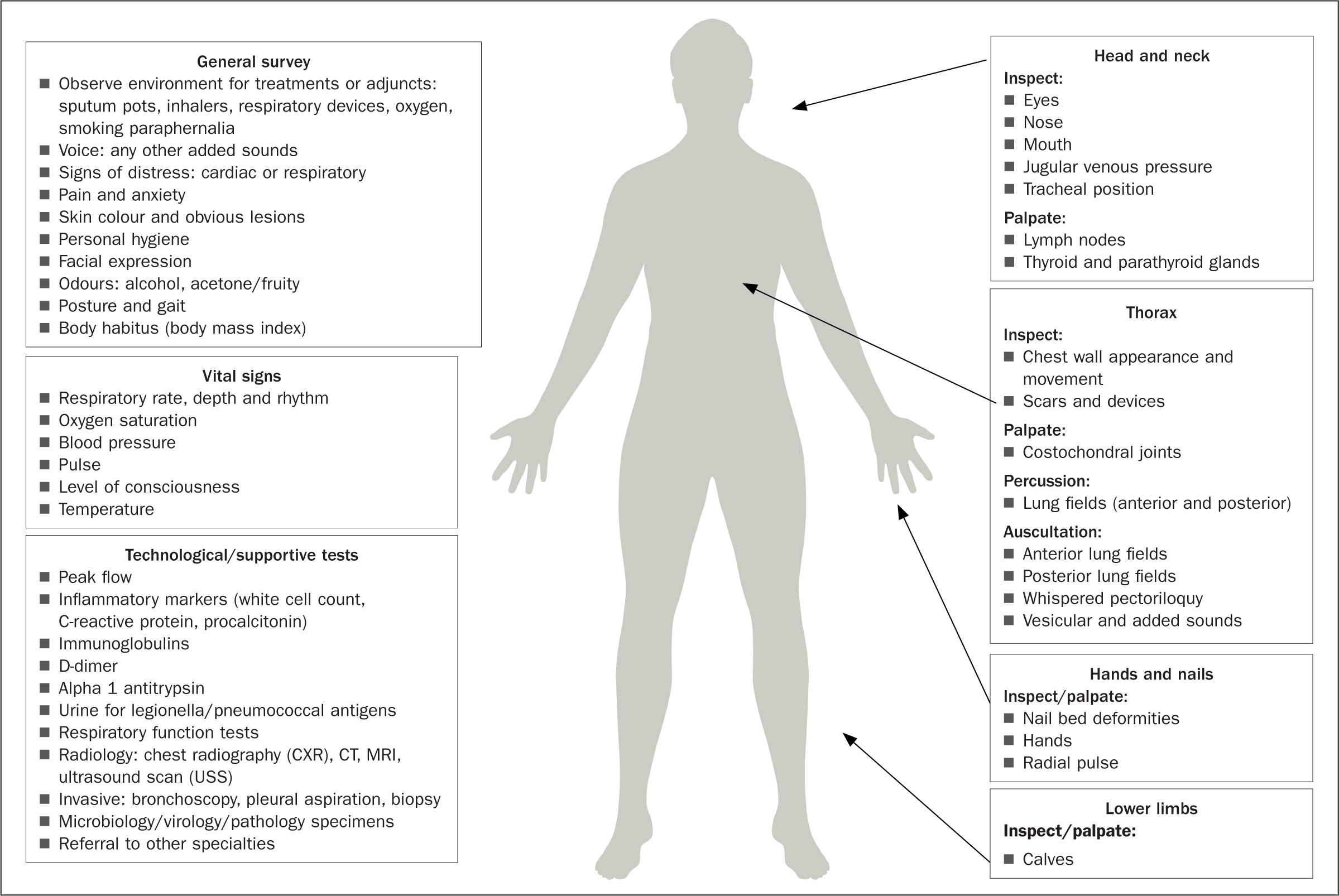
    The use of technology is particularly useful where diagnostic reasoning and stratifying disease severity are concerned. This then requires the ACP to possess essential skills of diagnostic interpretation, the fundamental principles of which are explored further by Roberts et al (2023). The focused respiratory examination, depicted in Figure 1, incorporates a traditional physical examination with common technological diagnostic testing.

    FIgure 1. Process for performing a respiratory examination (not exhaustive)

    Abnormal signs on physical examination

    Voice

  • Dysphonia (hoarseness) can be a sign of laryngitis
  • Stridor: laryngeal obstruction
  • Stertor: pharyngeal obstruction
  • Vocal fremitus: palpate the chest wall front and back while the patient says ‘ninety-nine’ aloud. Increased vibration over an area suggests tissue density (tumour, consolidation), decreased vibration over an area suggests fluid or air outside of the lung (pleural effusion or pneumothorax).
  • Face

  • Face – constriction of pupils and ptosis (Horner's syndrome from an apical lung cancer)
  • Mouth for central cyanosis, pursed lip breathing (chronic obstructive pulmonary disease (COPD)).
  • Hands/nails

  • Clubbing (thoracic disease; lung cancer, bronchiectasis, interstitial lung disease (ILD))
  • Peripheral cyanosis (hypoxia)
  • Tobacco (tar) staining (no filter rolled tobacco use)
  • Wasting of small muscles of the hand (lung cancer)
  • Tenderness at the wrist (hypertrophic pulmonary osteoarthropathy)
  • Flapping tremor – asterixis (carbon dioxide retention)
  • Fine tremor (beta 2 agonist use/overuse).
  • Neck

  • Accessory muscle use (COPD, respiratory muscle fatigue, airway obstruction)
  • Raised jugular venous pressure (JVP) (in cor pumonale)
  • Tracheal position – deviation can indicate shift in the mediastinum, pneumothorax, previous pneumonectomy.
  • Thorax

  • Chest shape abnormality (pectus excavatum, pectus carinatum, kyphoscoliosis, hyperinflated chest with intercostal indrawing)
  • Any scars evident (from previous surgery, trauma, chest drains)
  • Erythema and thickening of the skin (can be a result of previous radiotherapy)
  • Subcutaneous emphysema (crackling around the skin of the chest may indicate pneumothorax, bronchopleural fistula)
  • Asymmetry of chest movement (obstruction, infection)
  • Chest expansion – check for Hoover's sign over the xiphisternum, lack of expansion can support a diagnosis of COPD.
  • Breath sounds

  • Normal/vesicular – louder and longer on inspiration with no gap between inspiratory/expiratory sounds
  • Bronchial – similar sound to that heard over the trachea/larynx; breath sounds are equal in sound in both inspiratory/expiratory phase with a gap between evident. This is usually heard over areas of consolidation.
  • Added sounds

  • Wheeze: whistling sound (high pitched – asthma; low pitched – COPD)
  • Crackles: early inspiratory (COPD); late/pan inspiratory (alveoli-related disease) – may be:
  • Fine: typically ILD
  • Medium: typically left ventricular failure (LVF) or COPD
  • Coarse: typically pools of retained secretions ie bronchiectasis
  • Pleural friction rub – a creaking sound likened to treading in fresh snow. This occurs when thickened pleural surfaces rub together. It can indicate pleurisy, secondary to pulmonary infarction, pneumonia, pleural inflammation (systemic inflammatory disease or viral infection).
  • Tools and scoring systems

    In the presence of abnormal findings, clinicians may opt to use a risk-stratification tool or clinical scoring system. These are designed to present evidence to support decision making and treatment options as well as predicting outcomes and managing risk while improving efficiency (Dambha-Miller et al, 2020). It is pertinent to ensure any tools used have a strong evidence base supported by practice. Many have been quality checked through systematic review or other research process. Some commonly used systems/tools in respiratory diagnoses are as follows:

  • PERC Rule (Pulmonary Embolism Rule-out Criteria) to diagnose pulmonary embolism (Kline et al, 2008)
  • mMRC (Modified Medical Research Council) Dyspnoea Scale (Mahler and Wells, 1988) to measure breathlessness to diagnose the level of perceived respiratory disability
  • CRB65 score (NICE, 2016) to diagnose community-acquired pneumonia – mortality risk assessment
  • BCRSS (Brescia-COVID Respiratory Severity Scale) (Duca et al, 2020) to measure the severity of illness and level of care required for patients with COVID-19
  • GOLD criteria (Global Initiative for Chronic Obstructive Lung Disease) to measure chronic obstructive pulmonary disease severity (Agustí et al, 2023)
  • DRIP score (Drug Resistance in Pneumonia) to measure drug resistance in patients with pneumonia (Webb et al, 2016)
  • Wells criteria (Wells et al, 2001) to assess risk of pulmonary embolism
  • BODE index (Body-Mass Index, Airflow Obstruction, Dyspnoea, and Exercise Capacity Index) to assess COPD mortality. Includes a 6-minute walk test (Celli et al, 2004).
  • Conclusion

    This article has explored the consultation and clinical assessment skills required to perform a comprehensive history and clinical examination of those presenting with common respiratory pathology. Through clinical reasoning and effective communication, ACPs are often best placed to determine the nature of a patient's condition and focus on appropriate investigation techniques, which will guide the appropriate course of treatment.

    KEY POINTS

  • The role of the advanced clinical practitioner (ACP) has developed so that they are often the first contact for the patient
  • Developing the role to incorporate targeted clinical assessment is essential for accuracy, productivity and efficiency
  • Focused clinical assessments can save time, be more cost effective and, most importantly, ensure the patient gets the right treatment in a timely manner
  • Respiratory-focused assessment is complex; a good understanding of the evidence, and a systematic approach is essential for effective clinical practice
  • CPD reflective questions

  • What is the surface anatomy of the lungs?
  • What are the common causes of reduced chest expansion?
  • How might you differentiate consolidation and effusion clinically?
  • Do you understand the principles of evidence-based examination in the context of sensitivity and specificity of clinical examination findings?