

Lipodermatosclerosis (LDS), also referred to as sclerosing panniculitis, is a chronic skin and subcutaneous tissue condition arising from longstanding venous insufficiency (Bajor and Kotyla, 2024). The disorder often leads to a misdiagnosis of cellulitis, especially in its acute phase, due to overlapping signs such as pain, erythema and swelling (Klejtman et al, 2022). However, unlike cellulitis, which is an infectious process requiring antibiotics, LDS is an inflammatory condition that demands a multifaceted approach to treatment, focusing on reducing venous hypertension and addressing associated symptoms. Effective management of LDS requires timely recognition, proper differentiation from mimicking conditions and targeted therapy to prevent disease progression and optimise quality of life.
This clinical review explores the epidemiology, pathophysiology, clinical presentations (acute and chronic) of LDS, diagnostic considerations, differential diagnoses, management strategies, associated challenges and potential future directions of research for this condition. Due to the limited availability of recent studies and literature on LDS, older studies have been included to ensure a thorough evaluation.
Epidemiology
Over the years, various terms have been used to define this condition. In 1955, Huriez et al were the first to identify and describe LDS, initially referring to it as hypodermitis sclerodermiformis, and attributing its cause to cellulitis associated with venous insufficiency. Other terms, including sclerosing panniculitis and indurated cellulitis, have also been used to describe the condition. However, the terminology ‘lipodermatosclerosis’ has become the standard in both the USA and the UK (Miteva et al, 2010).
LDS is a manifestation of chronic venous disease (CVD), which varies in prevalence globally. In the UK, the Edinburgh Vein Study reported that approximately 9.4% of men and 6.6% of women demonstrate signs of CVD, with prevalence increasing with age (Tienthavorn, 2022). Although specific data on LDS prevalence are limited, it is estimated that about 10% of individuals with CVD develop LDS (Rabe et al, 2010). Globally, the prevalence of CVD ranges from 2.1% to 13.2%, depending on the population studied (Krishnan et al, 2020). Given that LDS is a complication of CVD, its occurrence is closely linked to the prevalence of venous insufficiency in different regions of the world. Further studies are required to explore the epidemiology of LDS, particularly its prevalence and incidence rates in both men and women, to provide a comprehensive understanding of its distribution and determinants.
Pathophysiology
The pathophysiology of LDS centres around chronic venous hypertension, which leads to impaired venous return and stasis (Kumar, 2022). Over time, increased venous pressure damages the microcirculation in the lower extremities, promoting inflammatory responses within the skin and subcutaneous tissues (Silverberg et al, 2023). Repeated inflammation results in fibrosis, which manifests as the characteristic induration and tightening of the skin in LDS.
Venous hypertension also triggers capillary leakage, allowing proteins and red blood cells to enter the interstitial space (Bilancini et al, 2021). This process contributes to chronic inflammation and the deposition of haemosiderin, leading to pigmentation changes often observed in chronic LDS. The underlying damage to the lymphatic system exacerbates fluid retention, further complicating the condition (Kumar, 2022).
Clinical presentation
Some studies have identified an association between LDS and obesity and, while it can occasionally appear bilaterally, it more commonly manifests unilaterally. LDS presents in two primary clinical forms: acute and chronic (Suehiro et al, 2023).
Acute LDS
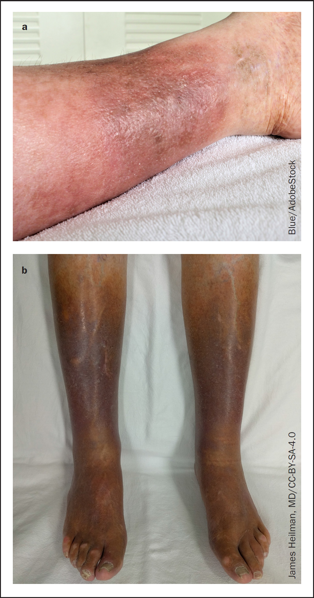
Acute LDS typically presents as a sudden onset of pain, erythema, and induration in the medial aspect of the lower leg, particularly in the gaiter region (DermNet, 2018; Osti et al, 2018) (see Figure 1a) The affected area is tender and inflamed, resembling cellulitis, but lacks systemic signs of infection, such as fever or lymphangitis. Swelling is usually absent or minimal, and the erythema is localised without spreading margins.
Chronic LDS
Chronic LDS involves the gradual development of fibrotic, woody plaques with tethering of the skin to underlying structures (see Figure 1b). The lower leg often develops an ‘inverted champagne bottle’ appearance due to progressive fibrosis, with a narrow ankle and widened upper calf (DermNet, 2018; Osti et al, 2018; Primary Care Dermatology Society, 2022). Additional signs of chronic venous insufficiency, such as hyperpigmentation, venous eczema, and ulceration, are frequently present.
Diagnosis
The diagnosis of LDS is primarily clinical, relying on a thorough patient history and physical examination (Bajor and Kotyla, 2024). Key diagnostic considerations include:
Patient history
Health professionals should take a comprehensive patient history to identify potential risk factors and underlying conditions linked to the development of LDS. Assessing for a history of venous insufficiency is critical, as this is the primary cause of LDS, leading to increased venous pressure and chronic inflammation. Inquiring about previous lower limb trauma or surgery is essential; these events can damage venous valves, exacerbating venous insufficiency. Similarly, a history of deep vein thrombosis (DVT) should be evaluated, as post-thrombotic syndrome can contribute to chronic venous insufficiency and subsequent LDS. Recurrent episodes of cellulitis should also be explored, as chronic inflammation and infection can worsen skin and subcutaneous tissue changes.
Peripheral vascular disease
Clinicians should inquire about a history of peripheral vascular disease and leg ulcers; these conditions can present with symptoms similar to LDS, such as skin changes, pain and swelling. Appropriate identification is crucial to differentiate LDS from these conditions, because the management strategies are different. Furthermore, when peripheral vascular disease or leg ulcers coexist with LDS, they can complicate the clinical picture by exacerbating tissue damage, impairing healing and increasing the risk of infections. Recognising these conditions allows for specific management plans that address all underlying problems.
Smoking and diabetes
Smoking status should be assessed because smoking is a significant risk factor for peripheral vascular disease, which can worsen tissue hypoxia and exacerbate LDS. Furthermore, clinicians should clarify whether patients have diabetes, because diabetes can impair wound healing and exacerbate chronic inflammation, further complicating the management of LDS.
Patient examination
Clinicians should perform a thorough neurovascular examination of the lower limbs when assessing a patient with suspected LDS.
Taking a thorough history ensures accurate diagnosis and informs effective management strategies (Alsararatee, 2024). Imaging studies, such as with venous Doppler ultrasound, can be useful to confirm venous insufficiency and exclude differential diagnoses, including DVT. A biopsy is rarely required but may be considered in atypical cases to rule out other causes of LDS or chronic skin changes. However, the American Osteopathic College of Dermatology (AOCD) (2025) has suggested that biopsy procedures may not be advisable in certain cases due to concerns regarding impaired wound healing and an increased risk of developing chronic complications. This recommendation highlights the importance of carefully evaluating the risks and benefits before undertaking invasive diagnostic interventions.
Differential diagnosis
LDS can be confused with several conditions, particularly cellulitis. Differentiating between these disorders is crucial to prevent unnecessary antibiotic use and ensure appropriate management. Common differential diagnoses include the following:
Management
The management of LDS focuses on addressing venous hypertension, controlling inflammation and alleviating symptoms. Key strategies include the following:
Patient education
Educating patients about the non-infectious nature of LDS is crucial (British Lymphology Society, 2022). Many patients receive repeated courses of antibiotics due to misdiagnosis as cellulitis, which is both unnecessary and potentially harmful. Emphasising the role of venous disease in the pathogenesis of LDS helps patients understand the rationale for treatment approaches, such as compression therapy and lifestyle modifications.
Treatment of underlying venous insufficiency
Addressing the root cause of venous hypertension may involve interventions such as endovenous ablation or sclerotherapy for varicose veins (Yao and Mukhdomi, 2023). Venous surgery in severe cases might be required (Reina-Gutierrez and Sanjuanbenito-Reina, 2023).
Compression therapy
Compression therapy is fundamental to LDS management (Rastel and Pichot, 2022). Graduated compression stockings (30–40mmHg) improve venous return, reduce oedema and prevent disease progression. It is essential to exclude peripheral arterial disease before initiating compression therapy. In acute LDS, compression may initially be poorly tolerated due to tenderness, so gradual implementation is recommended.
In randomised controlled trials, compression therapy using elastic compression stockings has been shown to effectively reduce skin induration in patients with hypodermitis. For instance, a study by Arendsen et al (2019) involving 17 patients with bilateral LDS, compared the effects of different compression stockings. In this study, one leg was treated with a stocking permanently impregnated with copper oxide ions, known for their biocidal, antimicrobial and wound-healing properties, whereas the other leg was treated with a non-impregnated stocking as a control. The findings showed that copper-impregnated stockings contributed to a reduction in the affected area of LDS.
Lifestyle modifications
Lifestyle modifications play a crucial role in managing risk factors for venous insufficiency and improving patient outcomes. Weight loss is particularly beneficial for overweight or obese individuals, because reducing body weight helps alleviate venous pressure and contributes to better overall outcomes (Australasian College of Dermatologists, 2019). Similarly, regular physical activity, such as walking, is also highly recommended. Exercise enhances venous return and reduces stasis, which is essential in mitigating the progression of venous insufficiency (AOCD, 2025). In addition, limb elevation during rest periods can significantly reduce swelling and discomfort in the affected leg, offering a simple yet effective strategy to manage symptoms (AOCD, 2025).
Pharmacological management
Analgesia is often necessary, particularly in acute LDS. Systemic anti-inflammatory treatments, such as corticosteroids, may provide short-term relief in acute cases, although their long-term efficacy is uncertain (Geist and Crane, 2023). Fibrinolytic agents have been suggested, but lack robust evidence to support routine use.
Management of chronic complications
Chronic LDS is often associated with venous ulcers, which require specialised wound care. Compression therapy remains the mainstay, supplemented by topical treatments and advanced wound dressings as required.
Challenges in management
The management of LDS encounters several challenges, despite advancements in understanding of the condition. Delayed diagnosis is a significant issue, because LDS is often misdiagnosed as cellulitis, leading to prolonged delays in initiating appropriate treatment and exacerbating disease progression. Another major challenge is patient adherence to recommended therapies, such as compression therapy and lifestyle modifications. Many patients struggle with compliance due to the discomfort and inconvenience associated with compression garments or a lack of understanding about the importance of these interventions (AOCD, 2025). In addition, there is limited evidence supporting the use of systemic treatments such as corticosteroids and fibrinolytic agents. Although these therapies show potential in managing LDS, their role remains a grey area, which restricts their broader application in clinical practice. Addressing these challenges is essential to improve outcomes and optimise the management of LDS.
Future directions
Research is required to explore novel therapeutic approaches for LDS, particularly systemic treatments that target inflammation and fibrosis. Studies evaluating the long-term efficacy of existing therapies, such as compression and corticosteroids, are also warranted. The integration of artificial intelligence (AI) in diagnostic processes presents an opportunity to enhance accuracy, particularly in distinguishing LDS from other similar conditions, helping to reduce misdiagnosis and unnecessary antibiotic use. Moreover, efforts to improve awareness among clinicians remain essential to optimise patient care outcomes, to reduce misdiagnosis and unnecessary treatments.
Conclusion
LDS is a complex condition that requires a holistic approach to diagnosis and management. Differentiating LDS from cellulitis and other mimicking conditions is essential to ensure appropriate treatment and avoid unnecessary interventions. Compression therapy, lifestyle modifications, and patient education form the foundation of effective management. Future research and enhanced clinical awareness will be essential in advancing the diagnosis, management and overall care of patients with LDS.