This website is intended for healthcare professionals
Search menu
References
Calpe-Damians N, Wennberg-Capellades L, Ventura-Rosado A Effectiveness and safety of a simple catheter securement device aimed at preventing catheterassociated urinary tract infection in intensive care unit patients: A randomized controlled trial. Nurs Crit Care. 2024; https://doi.org/10.1111/nicc.13111
Centers for Disease Control and Prevention. Catheter-associated urinary tract infection basics. 2024. https://tinyurl.com/ypt4hjum
Holroyd S Catheter valves: appropriate use and reduction of risk to bladder. Journal of Community Nursing. 2021; 35:(5)52-57
National Institute for Health and Care Excellence. Infection prevention and control: Quality statement 4—Urinary catheters. 2014. https://tinyurl.com/efevakws
Prieto J, Wilson J, Bak A A prevalence survey of patients with indwelling urinary catheters on district nursing caseloads in the United Kingdom: The Community Urinary Catheter Management (CCaMa) Study. J Infect Prev. 2020; 21:(4)129-135 https://doi.org/10.1177/1757177420901550
Royal College of Nurses. Catheter care: guidance for health care professionals. 2021. https://tinyurl.com/6zxsfe2m
Spoolder DAE, Geelhoed JP Management of bladder spasms in patients with indwelling urinary catheters: a systematic review. Continence. 2023; 7 https://doi.org/10.1016/j.cont.2023.100713
Urinary catheterisation is a crucial procedure in healthcare, requiring a thorough understanding of the anatomical and physiological differences between males and females to ensure safe and effective care. This guide explores the anatomy of the urinary system, detailing the kidneys, ureters, bladder, and urethra, and highlights key differences in catheterisation techniques for men and women. Emphasising evidence-based practices, it discusses recent advancements, such as antimicrobial catheters, and underscores the importance of comprehensive training and competency assessments for healthcare providers. Additionally, the guide advocates for patient-centred approaches, multidisciplinary collaboration, and adherence to updated policies to reduce complications such as catheter-associated urinary tract infections (CAUTIs). By integrating these critical evaluations and best practices, health professionals can enhance patient outcomes and maintain high standards of care in urinary catheterisation.
Urinary catheterisation is a critical procedure in health care for patients unable to naturally empty their bladders. It is essential that health professionals understand the anatomical and procedural differences between male and female catheterisation in order to ensure safe and effective care. This guide provides an updated overview of the urinary system's anatomy and detailed procedural steps for catheterisation in both men and women, supported by current best practices and recent research.
Anatomy of the urinary system
Male and female anatomy
A comprehensive understanding of the anatomy and physiology of the genitourinary tract in both genders is essential for effective catheterisation. This knowledge is crucial for health professionals, particularly nurses, to deliver holistic care and effectively communicate the procedure to patients. Understanding these anatomical differences also aids in anticipating potential complications and selecting the appropriate catheter size and type for each patient.
The urinary system
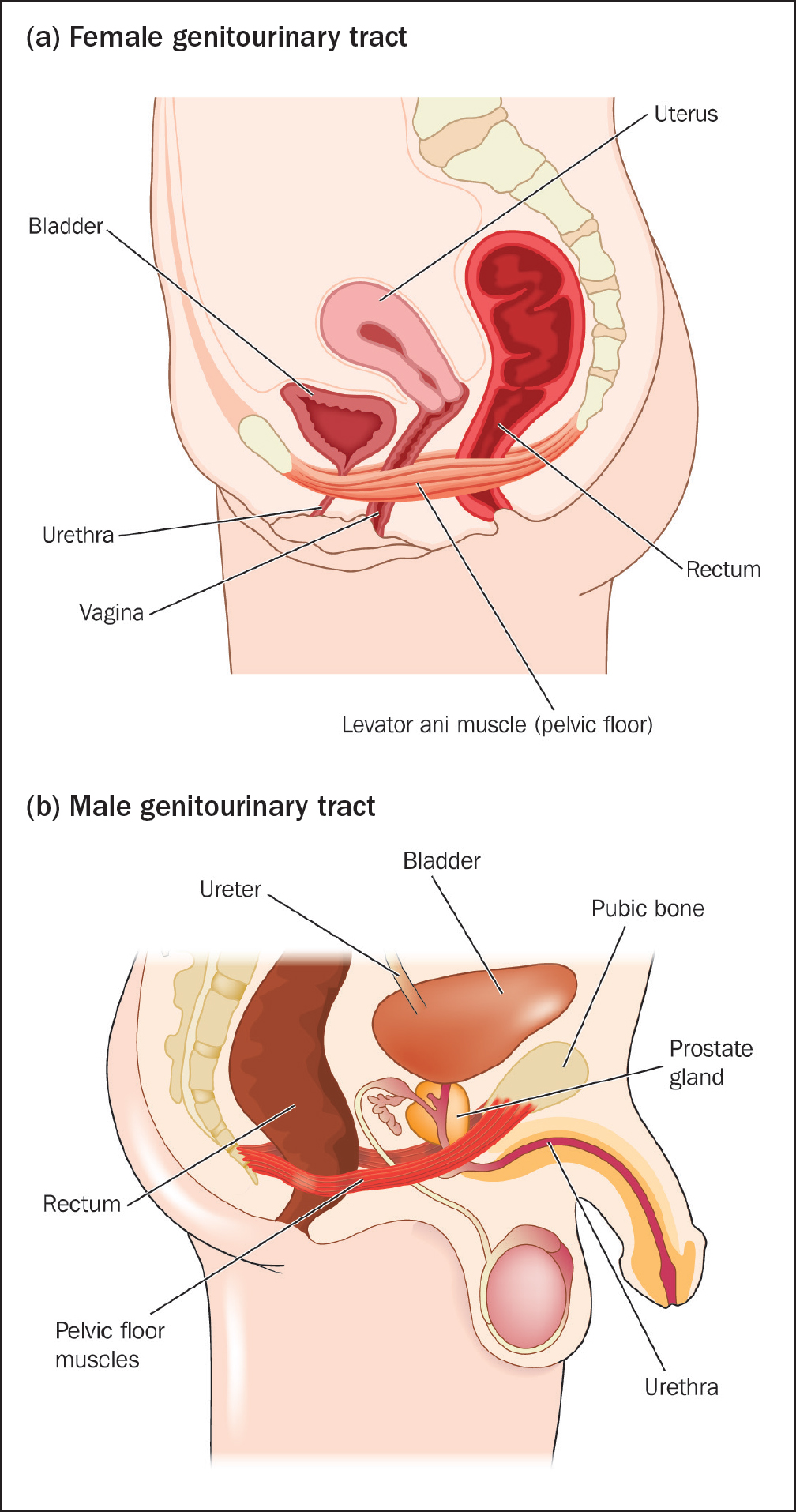
The urinary system, also known as the renal system, is responsible for the production, storage, and elimination of urine. It consists of the kidneys, ureters, bladder, and urethra (see Figure 1).
Kidneys: the kidneys are two bean-shaped organs located in the lower back. They filter waste products and excess fluids from the blood, forming urine. Each kidney contains approximately one million nephrons, the functional units that filter blood
Ureters: these are narrow tubes that carry urine from the kidneys to the bladder. Each kidney is connected to the bladder by a ureter. The smooth muscle walls of the ureters contract in waves to propel urine towards the bladder
Bladder: the bladder is a hollow, muscular organ that stores urine until it is ready to be expelled from the body. The bladder can expand to hold between 300 mL and 500mL of urine comfortably
Urethra: this tube carries urine from the bladder out of the body. In females, the urethra is approximately 3–4 cm long, whereas in males, it is about 20cm long. This significant difference in length necessitates different catheter designs and insertion techniques for men and women (National Institute for Health and Care Excellence (NICE), 2014).
Understanding the physiological processes of the urinary system, such as the filtration of blood by the kidneys and the storage capacity of the bladder, is equally important. This knowledge helps in assessing the patient's urinary function and identifying any abnormalities that may affect catheterisation.
Key anatomical differences in catheterisation
Female anatomy
The female urethra, which is 3–4cm long, is significantly shorter than the male urethra, and is located between the clitoris and the vaginal opening (Figure 1a)
The proximity of the female urethra to the vagina and rectum increases the risk of bacterial contamination and urinary tract infections, highlighting the need for meticulous aseptic techniques during catheterisation.
Male anatomy
The male urethra, which is approximately 20cm long, passes through the prostate gland and the length of the penis. This longer and more complex path requires a longer and often more rigid catheter (Figure 1b)
Men have an external sphincter muscle that controls the release of urine, located just below the prostate.
Importance of anatomical knowledge
This anatomical knowledge is vital for several reasons:
Holistic care: nurses can provide comprehensive care that considers the entire genitourinary system, addressing both physical and psychological aspects of patient health
Effective communication: clear and accurate explanations of the procedure can alleviate patient anxiety and improve co-operation during catheterisation
Procedural accuracy: understanding the anatomical differences and intricacies ensures that the correct catheter type and size are selected, and the procedure is performed correctly, reducing the risk of complications
Anticipation of complications: knowledge of the anatomical and physiological differences helps in anticipating and managing potential complications such as urethral trauma, incorrect catheter placement, and infections.
By integrating this detailed anatomical understanding into practice, health professionals can enhance patient outcomes, improve procedural success rates, and ensure a higher standard of care during urinary catheterisation.
Preparing for catheterisation
Nursing considerations
Nurses must operate within their competence limits, adhering to the Nursing and Midwifery Council (NMC) Code (NMC, 2018). Before catheterisation, it is essential to communicate with the patient, provide relevant information, and ensure the patient's understanding to obtain informed consent. Respecting the patient's privacy, cultural beliefs, and personal preferences is crucial. Box 1 illustrates the possible physical problems associated with catheterisation and Box 2 illustrates some key nursing considerations, in the form of the equipment that should be prepared for the procedure.
Increased risk of infection due to bacterial entry via the catheter
Allergies (lidocaine or latex): risk of allergic reactions ranging from mild irritation to severe anaphylaxis
Tissue damage: trauma to urinary tract tissues, potentially causing irritation or injury
Urethral strictures: scarring from repeated catheter use, leading to narrowing of the urethra
Bypassing and blockage: urine leakage or catheter obstruction due to improper size or debris formation
Equipment required for catheterisation
Sterile catheterisation kit: includes catheter, gloves, cleaning solution, lubricant, and drapes
Appropriately sized catheter: select correct size based on patient needs
Lubricant: sterile, water-based lubricant for easier insertion
Antiseptic solution: cleanse insertion site to reduce infection risk
Pre-filled anaesthetic gel syringe eg lidocaine
Sterile gloves: prevent contamination during the procedure
Drainage bag: ensure availability of sterile bag positioned below bladder level
Syringe with water: inflate catheter balloon for indwelling catheters
Protective pads: protect bedding and maintain a sterile environment
Step-by-step procedure for male catheterisation
Preparation: decontaminate hands, put on a plastic apron, and open the catheterisation pack using an aseptic nontouch technique (ANTT) (Royal College of Nursing (RCN), 2021)
Positioning: ask/assist the patient to retract his foreskin and, starting from the meatus, clean the glans in an upward direction using the gauze and saline. Clean the underside of the penis using a second piece of gauze in a single sweeping motion
Anaesthetic gel: after gaining consent, hold the penis in your non-dominant hand and use the dominant hand to insert the tip of the lidocaine gel syringe into the meatus, slowly squeezing the contents. Allow 5 minutes for the anaesthetic gel to take effect
Inserting the catheter: using both hands, place the sterile drape over the penis. Hold the penis in a fully extended position and use your dominant hand to insert the catheter into the urethral meatus. Advance the catheter slowly
Inflating the balloon: once urine starts to drain, inflate the balloon with 10mL of sterile water as per the manufacturer's instructions. Ensure the patient experiences no pain or discomfort
Securing the catheter: attach the catheter to the drainage bag and secure it to the patient's leg. Ensure the patient is comfortable and the area is clean and dry
Documentation: record the procedure immediately, including the date, time, type of catheter used, length, balloon size, volume of water inserted, batch number, manufacturer, expiry date, lubricant used, and any difficulties encountered (Box 3) (RCN, 2021).
Documentation
Date and time the catheterisation took place
The type of catheter used
The length of the catheter
Balloon size and the volume of water inserted
Batch number
Manufacturer
Expiry date
Lubricant used and expiry date
Document any difficulties/complications encountered on insertion
Name and signature of the health professional
Step-by-step procedure for female catheterisation
Preparation:decontaminate hands and open the catheterisation pack using ANTT (RCN, 2021)
Positioning: ask/assist the patient to lie in a supine position with legs bent and knees apart. Use a gauze swab to separate the labia and clean the urethral meatus using sterile saline
Anaesthetic gel: use the dominant hand to insert the tip of the lidocaine gel syringe into the meatus, slowly squeezing the contents. Allow 5 minutes for it to take effect (Geeky Medics, 2023)
Inserting the catheter: hold the labia open and insert the catheter into the urethral orifice in a backward and upward direction. Advance the catheter until urine starts to drain.
Inflating the balloon: inflate the balloon with 10mL of sterile water as per the manufacturer's instructions. Ensure the patient experiences no pain or discomfort
Securing the catheter: attach the catheter to the drainage bag and secure it to the patient's leg. Ensure the patient is comfortable and the area is clean and dry
Documentation: record the procedure immediately, including the date, time, type of catheter used, length, balloon size, volume of water inserted, batch number, manufacturer, expiry date, lubricant used, and any difficulties encountered (RCN, 2021).
Advanced considerations and complications
Catheter valves
Catheter valves are devices attached to the end of an indwelling urinary catheter that allow for controlled, intermittent drainage of urine. They support natural bladder filling and emptying cycles, which helps maintain bladder muscle tone and capacity, reducing the risk of bladder muscle atrophy. By promoting intermittent drainage, they also reduce the risk of urinary tract infections (Holroyd, 2021; Spoolder and Geelhoed, 2023). Catheter valves increase patient mobility and comfort, because they are more discreet and allow for greater freedom of movement compared with continuous drainage bags. This promotes a sense of independence and improves the quality of life for patients requiring long-term catheterisation. An overview of some of the positives and negative aspects of catheter valves is outlined in Table 1.
Aspect
Positives
Negatives
Infection control
Reduces the risk of urinary tract infections by allowing the bladder to fill and empty naturally
Improper use or poor hygiene can still lead to infections
Bladder function
Encourages normal bladder function and reduces the risk of bladder muscle atrophy
May not be suitable for all patients, particularly those with poor bladder control or cognitive impairments
Mobility
Increases patient mobility and comfort compared to continuous drainage
Requires more frequent emptying, which can be inconvenient for some patients
Convenience
Reduces the need for continuous drainage bags, which can be bulky and conspicuous
Patients need to remember to open and close the valve, which might be challenging for some individuals
Cost
Potentially lower costs over time due to reduced use of drainage bags and related supplies
Initial cost of valve may be higher than traditional catheters
Patient independence
Promotes independence and quality of life by reducing the stigma associated with visible urine bags
Requires patient education and compliance, which might not be feasible with all patients
Securement of catheters, tubing, and bags
The securement of catheters, tubing, and bags is crucial in preventing complications such as meatal tears in men. Various securement methods, including sleeves and straps, have been developed to enhance patient comfort and safety.
Securement devices
Sleeves: these provide a comfortable way to secure catheter tubing against the leg, reducing the risk of accidental tugging and subsequent trauma to the urethral meatus
Straps: soft, adjustable straps can be used to secure the catheter tubing to the thigh, offering a stable solution that minimises movement and potential for injury
Adhesive anchors: these devices stick to the skin and hold the catheter in place, reducing movement and the risk of dislodgment.
In a randomised controlled trial to assess the effectiveness of catheter securement, Calpe-Damians et al (2024) reported that proper securement of catheters can significantly reduce catheter-associated urinary tract infections (CAUTIs), the incidence of meatal tears and other catheter-associated complications.
Catheter passport
The‘catheter passport’ is a patient-held document that provides detailed information about their catheterisation. This includes the type of catheter used, the date of insertion, and any specific care instructions. The passport aims to improve communication between healthcare providers and patients, ensuring consistent and accurate care. The implementation of a catheter passport significantly reduced catheter-related complications and has been shown to improve patient satisfaction (Prieto et al, 2020).
Potential complications
Understanding potential complications is critical for effective catheterisation management. Complications can include CAUTIs, urethral damage, and patient discomfort (Centers for Disease Control and Prevention (CDC), 2024). It is essential to adhere to aseptic techniques and follow guidelines strictly to minimise these risks.
Strategies to minimise complications
Aseptic technique: always use sterile equipment and maintain ANTT during catheter insertion and handling (RCN, 2021)
Regular monitoring: monitor the catheter site regularly for signs of infection or complications. This includes checking for redness, swelling, or discharge (CDC, 2024)
Patient education: educate patients on catheter care, including hygiene practices and signs of potential complications. Empowering patients to monitor their condition can aid in the early detection of catheter-associated complications.
Conclusion
Urinary catheterisation is a crucial healthcare procedure requiring a thorough understanding of male and female anatomy, strict adherence to aseptic techniques, and proper equipment preparation. By following evidence-based practices and ensuring patient safety through careful communication and technique,healthcare providers can minimise complications and improve patient outcomes.
KEY POINTS
It is important to understand the anatomical differences between male and female urinary systems and their implications for catheterisation
Nurses must know how to perform safe and effective urinary catheterisation in both males and females, adhering to aseptic techniques
It is vital to identify and manage complications associated with urinary catheterisation using evidence-based practices
CPD reflective questions
How does your knowledge of the anatomical differences between male and female urinary systems shape your approach to urinary catheterisation in practice?
Which steps do you take to ensure the aseptic technique is maintained during catheterisation, and how do you assess your adherence to these practices?
How do you educate patients about urinary catheter care and the prevention of complications, and how could you improve your approach?
Reflect on a situation in which you encountered a complication during catheterisation. How did you address it, and what would you do differently to improve patient outcomes in future?